4. Imaging Tests Used in Lymphoma Diagnosis

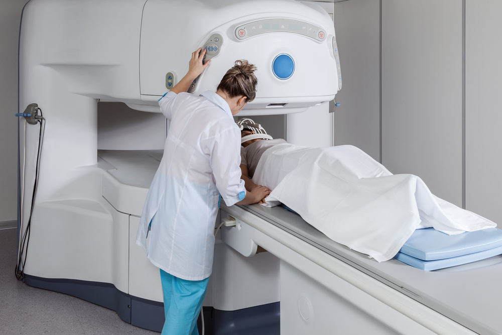
Imaging tests are conducted in patients suspected to suffer from lymphoma to examine the abnormal areas that may be affected by the disease. The most common tests are chest x-ray to search for swollen nodes in the chest, computed tomography (CT) scan that provides detailed images of the whole body including soft tissues, CT-guided needle biopsy used to drive the biopsy needle in suspicious areas, magnetic resonance imaging (MRI) scan to verify if the disease has spread to the brain or spinal cord, ultrasound to examine the damage in the internal organs and eventual existence of masses, positron emission tomography (PET) scans and gallium scans – both designed to determine the body parts with cancerous cells using radioactivity, or bone scan to understand the damages caused by lymphoma in the bones. In addition, the heart and lung function may also be tested to determine the accurate type of treatment.
Learn more about how PET scans can guide chemotherapy choices for Hodgkin’s lymphoma.
Lymphoma News Today is strictly a news and information website about the disease. It does not provide medical advice, diagnosis or treatment. This content is not intended to be a substitute for professional medical advice, diagnosis, or treatment. Always seek the advice of your physician or another qualified health provider with any questions you may have regarding a medical condition. Never disregard professional medical advice or delay in seeking it because of something you have read on this website.


