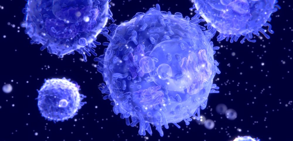
A new method for growing the T-cells used in lymphoma immunotherapies may some day lead to treatments that are faster and do a better job of honing in on a specific target.
A research team at two Harvard institutions developed a way to mimic signals from the antigen-presenting cells (APCs) that instruct T-cells to go after a target.
The team demonstrated that their method was faster and more effective than currently used methods for producing CAR T-cells and other cancer immunotherapies. The cells that the method generated were as good at fighting lymphoma in mice as those produced with current methods, researchers said.
“Prompted by recent breakthroughs in CAR T-cell therapies, we showed that a specific CAR T-cell product expanded with an APC-mimetic scaffold could facilitate treatment of a mouse model of a human lymphoma cancer,” Alexander Cheung, the first author of the study, said in a press release.
Cheung, who started the research at the Wyss Institute for Biologically Inspired Engineering and the John A. Paulson School of Engineering and Applied Sciences (SEAS), is now with UNUM Therapeutics.
Published in the journal Nature Biotechnology, the article, “Scaffolds that mimic antigen-presenting cells enable ex vivo expansion of primary T cells,” describes how the team built the scaffold, and the results of their experiments.
In the body, antigen-presenting cells instruct T-cells to attack a particular microbe or cancer cell. Researchers try to mimic the process using a tool called Dynabeads.
The beads are loaded with antibodies and factors that should prompt cells to target a cancer. Once T-cells are gathered from a patient, they are grown together with the beads in the laboratory, until there are enough specialized cells to infuse back into the patient.
But the team underscored that the method is not optimal. To start with, it is rather slow, taking weeks for enough cells to grow.
In addition, “the context in which these signals are presented is not representative of how they are naturally presented by APCs,” the researchers wrote, explaining that this can cause less than optimal expansion rates in addition to T-cells with limited or flawed functions.
Another problem is that the beads, which are not degradable, need to be separated from the cells before they can be used in a treatment, which further adds to the cost and time of the production.
Instead of beads, the team, led by Dr. David Mooney, developed rods made of mesoporous silica. They contain nano-sized pores that allow the entry of T-cells.
The rods, which assemble themselves into a three-dimensional scaffold, are first loaded with a T-cell activating factor. Then, scientists coat them with a layer of lipids, or fats, that resemble the outer membrane of antigen-presenting cells. Researchers then equip the rods with various antibodies, which are inserted into the membrane-like layer.
“Our approach closely mimics how APCs present their stimulating cues to primary T-cells on their outer membrane and how they release soluble factors that enhance the survival of the T-cells,” said Mooney, who is a faculty member at the Wyss Institute and leader of its Immunomaterials Platform. “As a result, we achieve much faster and greater expansion,” added Mooney, who is also the Robert P. Pinkas Family Professor of Bioengineering at SEAS.
The new system is also much more versatile, researchers said. By altering the composition of lipids and stimulatory factors, scientists can amplify only certain types of T-cells out of the mix of cells obtained from a blood sample.
Earlier studies show that defined subsets of T-cells, rather than a random mix, may be more effective when producing CAR T-cells.
“APC mimetic scaffolds enabled us to tune the ratios of subpopulations of T-cells with different roles in the desired immune responses, which in the future might increase their functionality,” said David Zhang, a graduate student working with Mooney who was a study co-author.
The scaffold was two to ten times more effective at expanding cells than Dynabeads, researchers said.
The team then produced CAR T-cells, again comparing the two methods. The next step was to inject the cells into mice with Burkitt’s lymphoma. This demonstrated that the cells performed similarly well, with similar safety, when produced by either of the two methods.
“The bioinspired T-cell-activating scaffolds, developed by the Wyss Institute’s Immunomaterials Platform, could accelerate the success of many immunotherapeutic approaches in the clinic, with life-saving impact on a broad range of patients, in addition to advancing personalized medicine,” concluded Dr. Donald Ingber, the Wyss Institute’s founding director.


